

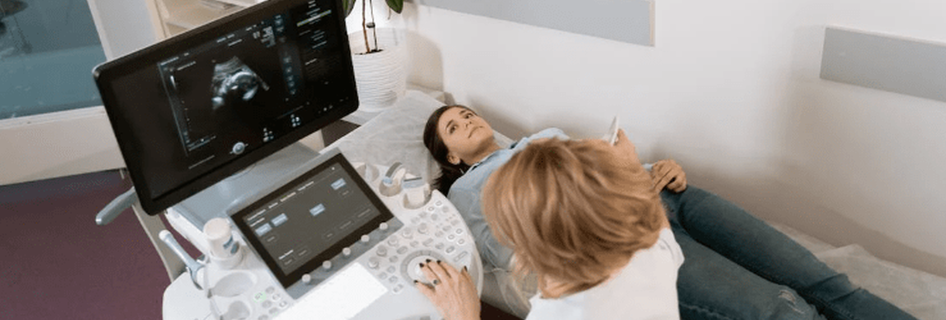
Gynecological ultrasound is an available, informative and safe method of diagnosing various women's problems.
The technique is used for preventive examinations and is actively used in the diagnosis of gynecological diseases. It is included in the list of mandatory examinations during pregnancy.
The diagnostic capabilities of the examination are enhanced by the use of expert-class equipment. The Universum clinic doctor is able to perform the procedure using all the necessary modes and make reasonable conclusions about the health of the woman and the fetus.
Indications for gynecological ultrasound
Gynecologists were among the first to appreciate the benefits of ultrasound. Currently, international standards of gynecological care define ultrasound diagnostics as the leading method of examination of women.
Indications for gynecological ultrasound include the following symptoms:
-
any pain in the pubic area and pelvis;
-
menstrual disorders (irregular menstruation, heavy, painful periods, bleeding during the intermenstrual period);
-
injuries of the perineum and pelvic organs;
-
appearance of tumors, protrusions in the groin, pelvic area;
-
uterine bleeding, discharge after menopause;
-
hormonal abnormalities (facial hair, male-type body shape changes);
-
infertility, absence of pregnancy after 6 months of regular attempts to conceive a child;
-
dyspareunia (discomfort and unpleasant sensations during sexual intercourse);
-
acute infections of the genitourinary system;
-
pathological discharge from the genitals (white);
-
a desire to install an intrauterine device or to monitor the condition of this contraceptive device;
-
discomfort and pain in the pelvis after gynecological operations.
The examination of teenagers and young girls is especially important. Ultrasound helps to assess the balance of sexual development and identify the cause of delayed or pre-mature puberty. No risk to the reproductive health of the pregnant woman.
Types of ultrasound in gynecology
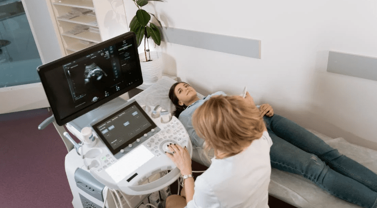
Initially, girls and women were examined "like everyone else", with the sensor applied to the abdominal skin. This technique is called transabdominal ultrasound. However, doctors soon realized that the technique was not very informative: approximately 30% of the pelvic volume remained inaccessible to ultrasound. For example, the cervix, cervical canal, and posterior pelvic cavity.
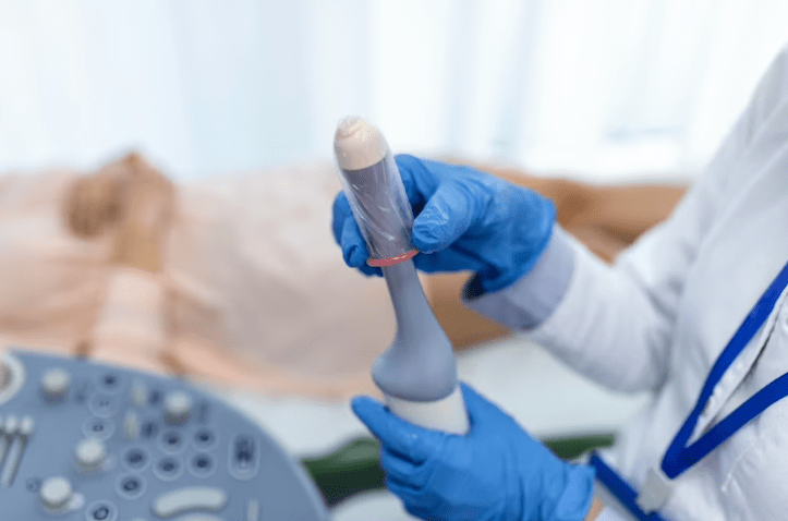
To correct this defect of ultrasound, gynecology needed a different approach: the transvaginal method. For this purpose, special cylindrical cavity sensors were developed. They are inserted into the vagina (and, if necessary, into the anus) and collect information about all organs and corners of the female pelvis.
The expert-class device used in Universum clinic operates in the mode that is necessary for the examination of the patient. To ensure the most complete examination, the clinic can use all types of gynecological ultrasound: transabdominal, transvaginal, and sometimes transrectal.
Limitations
The method is safe and widely used, even for the examination of pregnant women. However, sometimes gynecological ultrasound has to be abandoned. The main obstacle is acute infections of the abdominal cavity or vagina, which make it difficult for the transducer to come into close contact with the tissues.
After treatment and subsidence of the inflammatory process, the procedure can be performed without fear.
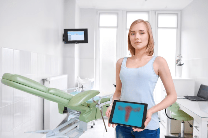
Diagnostic possibilities
The technological equipment and the experience of the clinic's diagnostician allow us to identify serious gynecological problems within one diagnostic procedure.
Gynecological ultrasound can be used to diagnose:
-
Polycystic ovary syndrome (PCOS);
-
ovulation and ovarian function disorders in adolescent girls, women of childbearing age and menopause
-
anatomical abnormalities in the structure of the internal genital organs;
-
tumor processes in the uterus (fibroids), appendages, pelvic tissue;
-
salpingo-oophoritis, endometritis;
-
polyps and cysts of the uterine cavity, adenomyosis, endometriosis;
-
prolapse of the internal genital organs (uterine prolapse);
-
ectopic pregnancy;
-
stenosis, deformation of the fallopian tubes (a common cause of female infertility);
-
purulent processes (e.g. pyosalpinx);
-
traumatic injuries to the reproductive organs, pelvic hemorrhage.
During the examination of a pregnant woman, the condition of the placenta, endometrium, amniotic fluid volume, and gestational age are assessed. It is possible to check whether the fetus is developing normally or whether there is a threat of abortion. And, of course, to find out the sex of the baby.

Due to the safety of ultrasound, the technique is used at all stages of medical care: from diagnostic examination to monitoring the treatment of a gynecological patient.
Special preparation for gynecological ultrasound
Air in the intestinal loops affects the diagnostic picture: it absorbs ultrasound waves. The day of the period when the examination is most informative is also important.
As a rule, preparation for gynecological ultrasonography involves, first of all, cleansing the intestines from gas. 2-3 days before the visit to the clinic, you should avoid foods that can cause flatulence: raw fruits and vegetables, milk, legumes and bread. Enterosorbents and simethicone-based antifoaming preparations may be useful.
As for the best time for the examination, the doctor at LuxMedic Clinic will suggest it: all women have a consultation with a gynecologist before a gynecological ultrasound.
Nothing more is required from the patient. Drinking water to fill the bladder is provided by the clinic. We also provide disposable condoms for the intravaginal gynecological ultrasound procedure, you do not need to bring them with you.
Benefits of gynecological ultrasound in Universum clinic
The accuracy of diagnosis depends on the equipment and the qualifications of the diagnostician. LuxMedic Clinic provides patients with both. The expert-class device allows you to perform ultrasound diagnostics in all modes: B-sonography, Doppler, 3D scanning, etc. The required mode is switched on from the sensor control panel and does not require any steps from the patient.
The clinic offers an ultrasound diagnostic service at home. We use a compact device with wide diagnostic capabilities.
A full range of sensors enables us to examine both women and girls. The procedure is carried out according to modern international protocols. As a rule, the conclusion with interpretation is ready on the day of the examination. In case of emergencies - immediately.
We guarantee affordable and informative gynecological ultrasound in Kyiv, Uzhhorod, Khmelnytskyi, Chernivtsi and Lviv – at any Universum clinic branch. Often, after a diagnostic procedure, there is no need for X-ray methods that are dangerous due to radiation exposure.
Sources
-
ISUOG Education Committee recommendations for basic training in obstetric and gynecological ultrasound. Ultrasound Obstet Gynecol. 2014 Jan;43(1):113-6. doi: 10.1002/uog.13208. Epub 2013 Nov 21. PMID: 24259320.
-
Silvestri MT et al. Frequency and Importance of Incomplete Screening Fetal Anatomic Sonography in Pregnancy. J Ultrasound Med. 2016 Dec;35(12):2665-2673. doi: 10.7863/ultra.16.01084. Epub 2016 Nov 7. PMID: 27821652.
-
Recker F et al. Point-of-care ultrasound in obstetrics and gynecology. Arch Gynecol Obstet. 2021 Apr;303(4):871-876. doi: 10.1007/s00404-021-05972-5. Epub 2021 Feb 8. PMID: 33558990; PMCID: PMC7985120.
-
Levine EM et al. Clinical Value of 3-Dimensional Ultrasound in Gynecology. J Ultrasound Med. 2018 Oct;37(10):2445-2450. doi: 10.1002/jum.14587. Epub 2018 Mar 2. PMID: 29498076.
-
Luczynska E, Zbigniew K. Diagnostic imaging in gynecology. Ginekol Pol. 2022;93(1):63-69. doi: 10.5603/GP.a2021.0209. Epub 2022 Jan 24. PMID: 35072254.
F. A. Q.
-
As a rule, you are invited for examination within the first 3-5 (maximum 10) days after menstruation, this is the follicular phase, the endometrial layer is thin and its abnormalities and uterine neoplasms are clearly visible;
-
if endometriosis is suspected, diagnostics is scheduled for other days, in the second half of the cycle;
-
if there is a problem with the ovaries, several sessions may be required - on day 6-7 of the menstrual cycle, on day 12-14 and after day 21;
-
women should first undergo a gynecological examination to choose the optimal period for the examination;
-
during pregnancy, diagnostics is carried out at different stages;
-
it is not recommended to plan diagnostics for the period of menstruation;
-
in case of symptoms of acute, dangerous conditions, gynecological ultrasound is performed immediately.
-
Eat food that can cause gas formation;
-
drink carbonated water and drinks;
-
have sexual intercourse with a man 48 hours before the procedure;
-
Empty the bladder an hour or more before the examination - the fluid in the bladder helps the doctor to orientate and recognise the pelvic organs;
-
before a transvaginal examination, on the contrary, the bladder is emptied, the doctor will ask the patient to do this, if necessary.
-
the product is put on the transducer for cavity examination when it is necessary to examine through the vagina or rectum;
-
condoms are disposable, if you need a transvaginal or transrectal scan, a new one is taken each time.
-
Nothing, everything you need for the procedure is provided by the clinic.


