


Breast ultrasound or ultrasound mammography is an affordable and highly informative method of non-invasive diagnosis of various forms of breast pathology. First of all, it is used to detect tumors and cancers in their initial stages, when they are easier to fight.

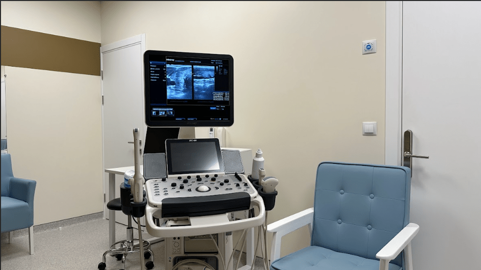
Thanks to the use of expert-class equipment, Universum clinic specialists diagnose focal and diffuse pathological processes in the mammary glands with high accuracy. Modern protocols of multiparametric ultrasound and evaluation of results according to the international US-BI-RADS system help in this.
Indications for Breast Ultrasound
All women between the ages of 18 and 40, even if they are in good health, will benefit from an annual breast ultrasound. After the age of 40, after the menopause, it is worthwhile to be examined twice a year. The safety of ultrasound allows this.
You should visit the clinic for a special ultrasound of the mammary glands if you have pathological symptoms in the breast
-
discharge from the nipples, even sparse, visible only on the underwear
-
change in the shape or volume of one breast, asymmetry
-
severe lumping (thickening), tenderness and swelling of the breast before menstruation;
-
pain in any part of the breast at any time during the cycle;
-
chronic diseases of the reproductive system (ovaries, uterus);
-
lumps in the breast tissue that can be palpated or felt with the fingers;
-
nipple deformation, retraction;
-
focal or generalized redness of the breast skin;
-
changes in skin texture (infiltration, induration);
-
discomfort and pain following chest trauma;
-
monitoring the condition of breast implants.
The examination is performed on adolescent girls if they have hormonal imbalances. The procedure is not contraindicated for pregnant women or women who are breastfeeding.
Surprisingly, men also need to undergo an ultrasound examination of the mammary glands. In exceptional cases, they may also need help due to the development of breast cancer, various forms of mastopathy.
Diagnostic value
The equipment for thoracic ultrasound at LuxMedic Clinic allows to perform diagnostics in all necessary modes (grayscale sonography, elastography, Doppler, creation of a 3D model of the examined organ).
The wide range of functions of the device makes it possible to detect various diseases and pathological processes in breast tissue:
-
the degree of fibrous changes in the breast parenchyma;
-
fibroadenomas, cysts, malignant nodes;
-
various forms of mastopathy (diffuse, nodular, fibrocystic);
-
diffuse changes (malignant or benign)
-
swelling, abscesses, inflammation in mastitis;
-
post-traumatic hematomas;
-
lactation disorders (lactostasis, calcification, galactoceles);
-
characteristics of the milk ducts (intraductal papillomas, tumors);
-
condition of regional lymph nodes;
If necessary, we use ultrasound guidance during the biopsy or puncture procedure.
Echography is particularly valuable for visualizing tissues near the sternum. These areas are poorly differentiated by radiographic methods.
In addition, only Doppler ultrasound of the mammary glands provides a picture of the blood flow in the area being examined. These features allow malignant tumors to be distinguished from benign ones. The use of safe contrast agents for ultrasound helps to improve diagnostic data.
Contraindications
The technique is uniquely safe. It can be performed on patients of any age, as well as on pregnant and breastfeeding women.
Active infection of the area to be examined may force you to postpone the procedure. After the doctor cures the inflammatory process, the breast ultrasound can be performed in full.
Problems may also arise during the examination of small children, who are difficult to hold in place, but in cooperation with the parents, the specialists of the LuxMedic Clinic manage to solve this problem.
Preparation
None is required. Ultrasonography of the mammary glands can be performed on the day of the visit. In order to obtain accurate diagnostic data, the clinic's mammologist may prescribe the procedure for different periods of the menstrual cycle.

As a rule, it is better to perform the examination in the first half of the cycle, before ovulation. It is better to contact the clinic's consultant in advance and specify the optimal time for the procedure.
Procedure
In the diagnostic room, a woman takes off her clothes and bra and lies down on the couch. The optimal position is on the back; if the mammary glands are large, the doctor may ask you to lie on your side.
A layer of gel is applied to the skin of the area to be examined to improve the conductivity of the ultrasound waves. The physician places the transducer on the body and examines the ultrasound image on the screen of the device. The diagnostic modes are switched from the control panel of the device and the woman does not have to do anything.
When the diagnosis is complete, the gel residue is wiped dry with a disposable towel. The woman dresses and returns to her normal routine.
References
-
Guo R, Lu G, Qin B, Fei B. Ultrasound Imaging Technologies for Breast Cancer Detection and Management: A Review. Ultrasound Med Biol. 2018 Jan;44(1):37-70. doi: 10.1016/j.ultrasmedbio.2017.09.012. Epub 2017 Oct 26. PMID: 29107353; PMCID: PMC6169997.
-
Bhatt AA, Whaley DH, Lee CU. Ultrasound-Guided Breast Biopsies: Basic and New Techniques. J Ultrasound Med. 2021 Jul;40(7):1427-1443. doi: 10.1002/jum.15517. Epub 2020 Sep 30. PMID: 32997819; PMCID: PMC8246574.
-
Smith DN. Breast ultrasound. Radiol Clin North Am. 2001 May;39(3):485-97. doi: 10.1016/s0033-8389(05)70293-1. PMID: 11506089.
-
Covington MF. Ultrasound Elastography May Better Characterize BI-RADS 3 and BI-RADS 4A Lesions to Decrease False-Positive Breast Biopsy Rates and Enable Earlier Detection of Breast Cancer. J Am Coll Radiol. 2022 May;19(5):635-636. doi: 10.1016/j.jacr.2022.02.023. Epub 2022 Mar 19. PMID: 35317989.
-
Tang CY, Guan PS, You QQ, Yuan HX, Wang WP. Contrast-enhanced ultrasound combined with ultrasonic elastography to diagnose encapsulated papillary carcinoma: A case report. Clin Hemorheol Microcirc. 2022;82(4):391-396. doi: 10.3233/CH-221558. PMID: 36057816.
F. A. Q.
The breast is swollen, deformed, and the nipple is retracted;
-
There is asymmetry of the breast glands;
-
A lump of any shape, mobility and sensitivity is felt in the breast;
-
The breast is reddened, swollen and pathological discharge (mucus, blood) from the nipple is noted;
-
A nursing mother has lost milk, the breast is swollen and painful;
-
There has been an injury to the breast;
-
Chest pain is a reason to get checked out;
-
Even if you are in perfect health, you should have a yearly exam from the age of 18;
-
From the age of 40, a screening is recommended every 6 months.
- No special preparation is necessary;
- You just need to contact the clinic's consultant and make an appointment;
- Everything you need for the procedure will be provided by the clinic.
- Correspondence of the development of the glands with the patient's age;
- infectious processes (mastitis, abscess, inflammation of the ducts)
- Any form of mastopathy;
- Fibroadenomas, adenomas, cysts;
- Pathology of the ducts (deformations, intraductal papillomas, tumors);
- hematomas in glandular tissues and under them, in muscles;
- Condition of lymph nodes;
- Degree of fibrous changes;
- Diagnostic conclusion according to the US-BI-RADS protocol allows to assess the risk of malignant tumor development in the scanned organ.
- There are no specific requirements;
- It is useful to inform the doctor if the patient has implants.


