

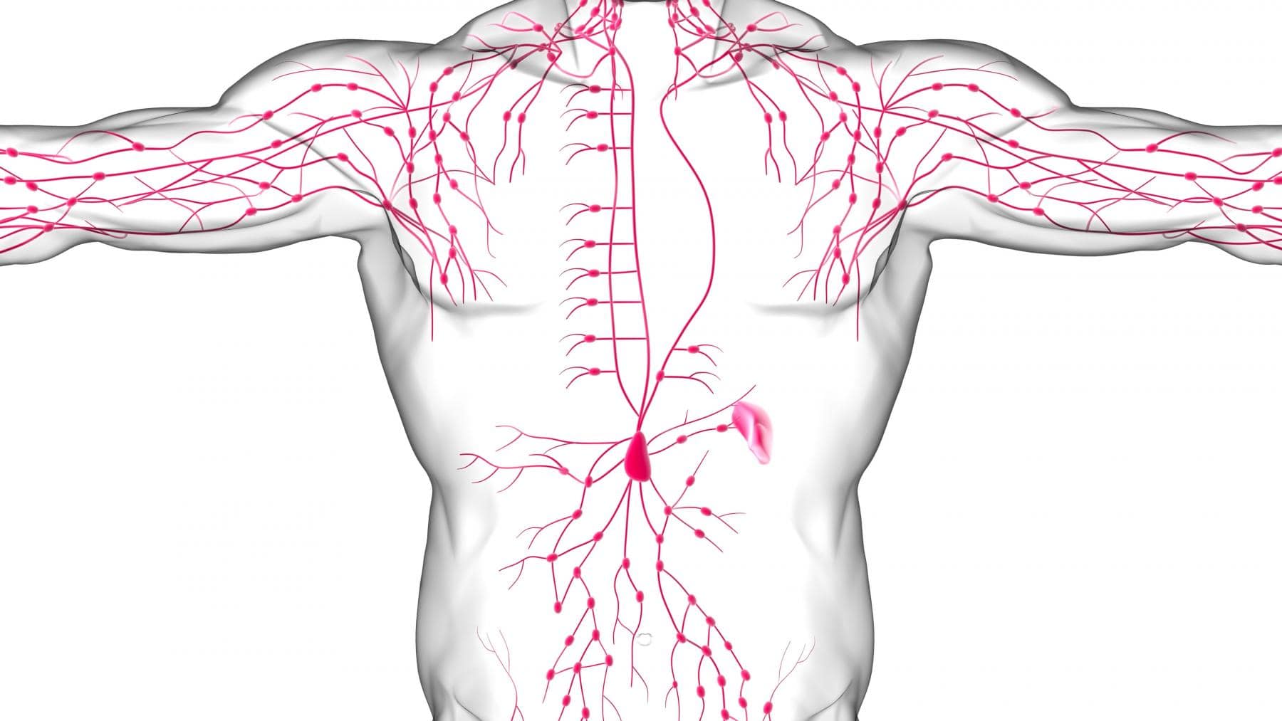
Lymph node ultrasound is a non-invasive technique for diagnosing the peripheral lymphatic system. It allows you to determine what diseases the patient has, and confirm the diagnosis. Note that lymph nodes are located practically throughout the body. But ultrasound of lymph nodes is carried out only in certain areas.
The examination is subjected to:
- cervical;
- occipital;
- auricular;
- submandibular;
- axillary;
- popliteal;
- retroperitoneal nodes.
Organs located in the groin may also be examined.
Indications for ultrasound of lymph nodes
With various diseases, patients can develop soreness, thickening or enlargement of lymph nodes. Usually this occurs due to the course of inflammatory processes, in metabolic disorders, etc. Let's consider in what cases diagnosis is prescribed:
- Ultrasound of the lymph nodes of the neck is carried out if the patient has complications of ARI, suspected laryngitis, pharyngitis, rubella, sore throat, diphtheria and other diseases.
- The examination of the nodes on the lower jaw is carried out if there is a suspicion of leukemia or lymphadenitis.
- Ultrasound of inguinal lymph nodes is performed if the patient often has pain in the legs and lower abdomen, as well as with problems with pelvic organs.
The retroperitoneal nodes are checked if there are suspicions of cancer or metastases. In any case, the decision to conduct diagnostics is made by a doctor. Note that ultrasound is carried out in conjunction with other diagnostic measures. The study allows you to confirm the diagnosis, not to make it.
Contraindications to ultrasound of lymph nodes
This procedure is considered completely safe. It can be performed even in children or pregnant women. There are temporary contraindications in the form of open wounds or skin damage in the necessary places. After healing, you can conduct a full-fledged diagnosis.
How to prepare for the procedure
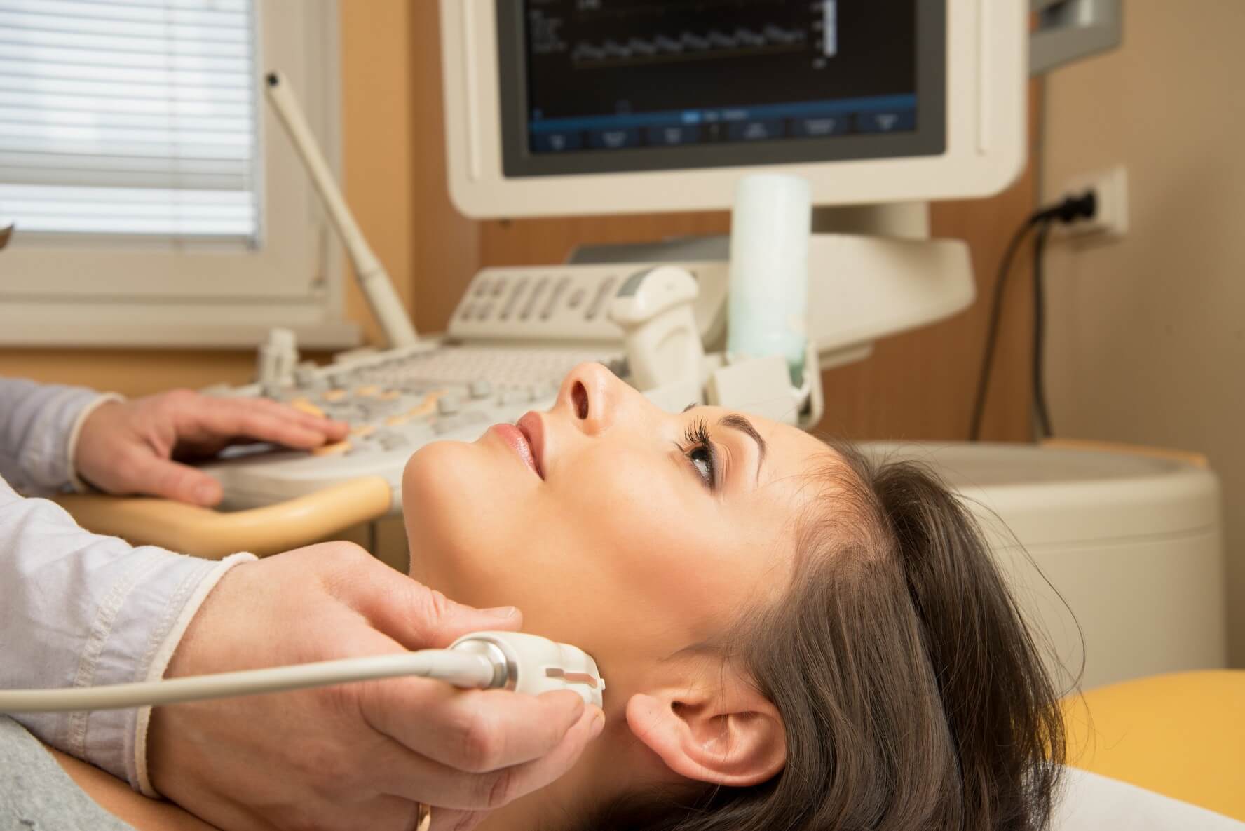
Since the procedure is completely safe, the patient does not need to prepare for it in any way. Except to take a bath the day before so that the skin is clean. Ultrasound is usually performed in a lying or sitting position (it all depends on what is being checked). If the size of the lymph nodes is enlarged, the patient may feel discomfort or pain when touching the sensor. The procedure itself takes about 15-20 minutes, after which you can get the result and use it for further diagnosis.
What the study may reveal
Making ultrasound lymph nodes in Kiev, you can determine the presence of inflammatory processes, find malignant tumors, various mycoses and lymphoma. Also this method allows you to determine the presence of syphilis or tuberculosis.
Literature
- ncbi.nlm.nih.gov/pmc/articles/PMC2324368/
- sciencedirect.com/science/article/pii/S0887217117300586
- en.wikipedia.org/wiki/Lymphadenopathy
- healthimages.com/what-do-swollen-lymph-nodes-mean
F. A. Q.
This procedure allows you to confirm the diagnosis made by the doctor, or clarify it. It can also be used to assess their localization, number, structure and shape.
During the examination, you can see the location and size of the nodes, the presence of neoplasms in them, the passage of blood flow through them and the general condition of the organs.
If you have cancer, the nodes will begin to increase in size. They filter and destroy cancer cells, which is why inflammation occurs. But they can not always cope with the disease. Therefore, if the first symptoms of inflammation appear, you should contact the clinic.
In a healthy person, only three types of nodes can be felt: submandibular, inguinal and axillary. Their size should not exceed 10 mm. If inflammation begins, they increase. In case of metastatic lesions, the neoplasms themselves can be felt.

